
Tissue Growth and Developmental Pattern Formation
Biology

Anna Kicheva
Professor
1 Career
- Since 2024 Professor, Institute of Science and Technology Austria (ISTA)
- 2015 – 2024 Assistant Professor, Institute of Science and Technology Austria (ISTA)
- 2008 – 2015 Postdoctoral fellow, MRC-NIMR (Francis Crick Institute), London, UK
- 2003 – 2008 PhD, University of Geneva, Biochemistry Dept., Switzerland (2006-2008) Max Planck Institute of Cell Biology and Genetics, Dresden, Germany (2003-2006)
- 2000 – 2003 B.A., Bennington College, Bennington, VT, USA
1.1 Selected Distinctions
- Starting 2028 Member of Scientific Advisory Board of Collaborative Research Center “Neurodrive” (Erlangen, Germany), led by Chichung Lie and Marissa Karow
- 2027 Chair of 2027 Developmental Biology Gordon Research Conference.
- 2026 ANR Priority Research Program and Equipment international grant panel member
- 2025 NIH/NICHD site visit evaluation panel member
- Since 2022 Editorial Board of PLOS Biology
1.2 Selected Grants
- 2022 – 2027 ERC Consolidator Grant, Mechanisms of tissue size regulation in spinal cord development, € 1,993,094.
- 2024 – 2028 Special Research Program (SFB) grant part II, FWF Austria, Neural stem cell modulation, Kicheva group awarded ~ € 400,000.
- 2016 – 2021 ERC Starting Grant, Coordination of patterning and growth in the spinal cord, € 1,499,119.
1.3 Tech Transfer (Patents, Spin-Offs)
N/A
1.4 Selected Publications
- Lehr S§, Brückner DB§, Minchington T, Greunz-Schindler M, Merrin J, Hannezo E*, Kicheva A*. (2025) Self-organised pattern formation in the developing neural tube by a temporal relay of BMP signalling. Developmental Cell 60(4), 567-580. Available: https://doi.org/10.1016/j.devcel.2024.10.024.
- Lehr S§, Merrin J§, Kulig M, Minchington T, Kicheva A*. (2024)Fabrication of elastomeric stencils for patterned stem cell differentiation. STAR Protocols 5 (4) 103187. Available: https://doi.org/10.1016/j.xpro.2024.103187.
- Ho R§, Kishi K§, Majka M, Kicheva A*, Zagorski M*. (2024) Dynamics of morphogen source formation in a growing tissue. PLOS Comput Biol. Available: https://doi.org/10.1371/journal.pcbi.1012508.
- Bocanegra-Moreno L, Singh A, Hannezo E, Zagorski M*, Kicheva A*. (2023) Cell cycle dynamics controls fluidity of the developing mouse neuroepithelium. Nature Phys 19, 1050–1058. Available: https://doi.org/10.1038/s41567-023-01977-w.
- Guerrero P, Perez-Carrasco R, Zagorski M, Page D, Kicheva A*, Briscoe J*, Page K*. (2019) Neuronal differentiation affects tissue mechanics and progenitor arrangement in the vertebrate neuroepithelium. Development 46(23). pii: dev176297. Available: https://doi.org/10.1242/dev.176297.
2 Group Members
- Paraskevi Athanasouli, Postdoc since 2025
- Nursen Balekoglu, Postdoc since 2025
- Rohit Krishnan Harish, Postdoc since 2022
- Kasumi Kishi, Postdoc since 2025
- Corina Luca, Postdoc since 2025
- Amrita Singh, Postdoc since 2021
- Tobias Viehböck, Research Technician since 2026
3 Research
Every complex organism begins as a single cell, yet reliably forms organs with precise sizes and shapes. How such reproducibility emerges from inherently noisy biological processes remains a central open question in biology. At its core is the interplay between morphogen signaling, tissue growth, and cell fate specification, which together generate emergent properties such as size, scaling, and patterning precision. However, which interactions are critical and how they ensure robust outcomes remain largely unknown. My lab addresses this challenge by combining quantitative experiments with multiscale modeling in the developing vertebrate central nervous system, focusing on the spinal cord as a tractable and medically relevant model.
Our work spans three main areas. First, we study how morphogen gradients are established and controlled. We showed that key signaling domains form through biphasic dynamics that separate early specification from later expansion, enabling robust cell type generation and scaling with tissue size. Second, we investigate how cells interpret these signals. We found that cells integrate multiple, often opposing inputs within shared gene regulatory networks to extract positional information with high precision, a likely general principle in development. Third, we examine how tissue growth feeds back on patterning. Combining experiments with mechanical modeling, we showed that growth rate influences tissue shape, material properties, and patterning precision.
Together, this work establishes a quantitative framework for how signaling, growth, and mechanics are coordinated to ensure reproducible development, with broad relevance for organ formation, disease, and regenerative medicine.
3.1 Current Scientific Projects/Directions
- Regulation of restorative growth in the neural tube
- Cross-talk between morphogen gradients
- Regulation of roof plate specification and size
- BMP gradient formation
- Notochord morphogenesis
- Morphogen-controlled gene regulatory networks in patterning and growth
3.2 Research in Focus
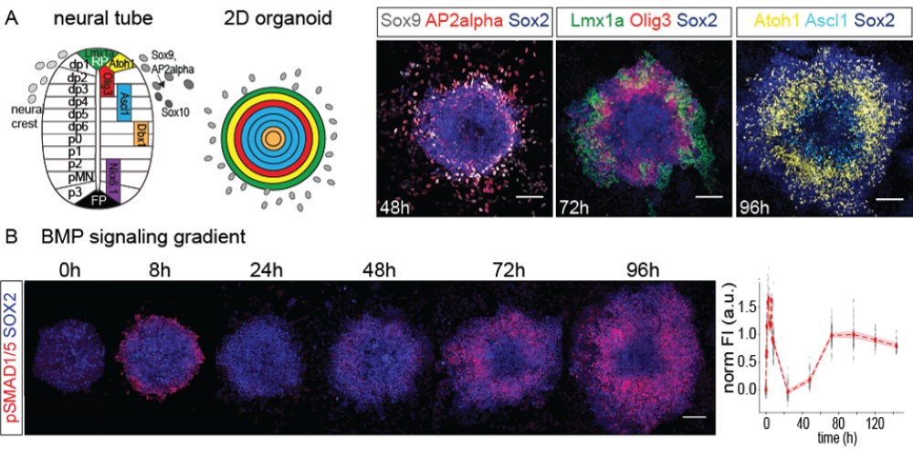
B. A BMP signaling gradient (red) is rapidly established, subsequently downregulated, and later activated again. These dynamic and reproducible changes in signaling activity guide the proper establishment of spatial patterning
In the dorsal neural tube, parallel gradients of BMP and Wnt signaling control pattern and growth, but it is unclear how these gradients are established and interpreted. To address this, we developed a novel 2D stem cell differentiation system based on stencil micropatterning. Unlike traditional micropatterning approaches, where cells are cultured on confined surfaces, our system allows unconstrained colony growth and cell migration. This allows us to achieve simultaneous standardized quantitative readouts of signaling, pattern formation, and colony growth.
Remarkably, in this system, dorsal neural tube pattern self-organizes into nested concentric domains. We found that pattern formation depends on the emergence of BMP and Wnt signaling gradients with characteristic biphasic temporal dynamics. In collaboration with the Hannezo group at ISTA, we developed a data-driven theoretical framework and showed that these biphasic dynamics are produced by a temporal relay mechanism that couples fast inhibition and slow positive regulation of BMP signaling. The inhibition is mediated by the complementary activity of several BMP inhibitors, while the activation depends on the specification of an endogenous source of BMP production by Lmx1a and Wnt. We observed similar dynamics of BMP signaling in vivo. The temporal relay mechanism that we uncovered allows the specification of distinct cell fates (neural plate border and neural progenitors) at defined developmental times.
Altogether, in this work, we identified a novel mechanism that underlies the temporal dynamics of BMP signaling and organizes pattern formation in the dorsal neural tube. Our findings show how morphogen gradients can self-regulate their shapes by inducing target genes that control morphogen production. We also developed a new 2D stem cell stencil differentiation system that provides new opportunities for quantitative studies of pattern formation and growth control. This work was published in Lehr et al, Developmental Cell 60(4), 567-580 (2025) and Lehr et al, STAR Protocols 5 (4) 103187 (2024).

