
Virus, Cell and Tissue Architecture
Biology

Florian Schur
Professor
1 Career
- Since 2024 Professor, Institute of Science and Technology Austria (ISTA)
- 2017 – 2024 Assistant Professor, Institute of Science and Technology Austria (ISTA)
- 2016 – 2017 Bridging Postdoctoral Researcher, European Molecular Biology Laboratory (EMBL), Heidelberg, Germany
- 2012 – 2016 PhD, EMBL Heidelberg, Germany
1.1 Selected Distinctions
- 2023 ERC Starting Grant
- 2021 FEBS Excellence Award
- 2021 EMBO Young Investigator
1.2 Selected Grants
- 2024 – 2029 AustrianScienceFund(FWF)EmergingFieldsgrant; “Crucial Steps in Evolution: The rise of genome architecture”; Co-Investigators: Frédéric Berger (GMI Vienna) and Christa Schleper (University of Vienna); Awarded overall: € 4.4 million, awarded to RG: € 1.3 million.
- 2023 – 2028 ERC Starting Grant; “ActinID: A molecular atlas of Actin filament IDentities in the cell motility machinery”; GrantNo. 101076260; awarded to RG: € 1,499,531.
- 2021 – 2025 Chan Zuckerberg Initiative Frontiers of Imaging Program; “CryoMinflux-guided in-situ visual proteomics and structure determination”; Co-Investigator Johann Danzl; Grant No. DAF2021234754; Awarded overall: € 835,000; Awarded to RG: € 417,000.
1.3 Tech Transfer (Patents, Spin-Offs)
N/A
1.4 Selected Publications
- Carpentier R, Kim J, Capizzi M, Kim H, Fäßler F, Hansen JM, Kim MJ, Denarier E, Blot E, Arnal I, Marcaida MJ, Dal Peraro M, Kim D, Schur FKM, Song JJ, Humbert S. Structure of the Huntingtin F-actin complex reveals its role in cytoskeleton organization. Science Advances (2025), https://doi.org/10.1126/sciadv.adw4124.
- Obr M, Percipalle M, Chernikova D, Yang H, Thader A, Pinke G, Porley D, Mansky LM, Dick RA, Schur FKM. Distinct stabilization of the human T-cell leukemia virus type 1 immature Gag lattice. Nature Structural and Molecular Biology (2024), https://doi.org/10.1038/s41594-024-01390-8.
- Zens B, Fäßler F, Hansen JM, Hauschild R, Datler J, Hodirnau V-V, Zheden V, Alanko J, Sixt M, Schur FKM. Lift-out cryo-FIBSEM and cryo-ET reveal the ultrastructural landscape of extracellular matrix. Journal of Cell Biology (2024), https://doi.org/10.1083/jcb.202309125.
- Datler J, Hansen JM, Thader A, Schlögl A, Bauer L, Hodirnau VV, Schur FKM. Multi-modal cryo-EM reveals trimers of protein A10 to form the palisade layer in poxvirus cores. Nature Structural and Molecular Biology (2024), DOI: 10.1038/s41594-023-01201-6.
- Fäßler F, Javoor MG, Datler J, Döring H, Hofer FW, Dimchev G, Hodirnau VV, Faix J, Rottner K, Schur FKM. ArpC5 isoforms regulate Arp2/3 complex-dependent protrusion through differential Ena/VASP positioning. Science Advances (2023), DOI: 10.1126/sciadv.add6495.
2 Group Members
- Darya Chernikova, PhD Student since 2022
- Manjunath Javoor, PhD Student since 2021
- Mathias Percipalle, PhD Student since 2022
- Noah Weber, PhD Student since 2025
- Benoit Arragain, Postdoc since 2025
- Miroslav Homola, Postdoc since 2024
- Seungha Hwang, Postdoc since 2025
- Kain van den Elsen, Postdoc since 2023
- Andreas Thader, Research Technician since 2017
3 Research
Numerous biological properties are governed by architectural and structural principles that depend on the higher-order, three-dimensional organization of biomolecules. This is manifested, for example, in the cytoskeleton, extracellular matrix, genome architecture, and viral assembly.
How this organization is established, maintained, and dynamically adapted remains incompletely understood, largely due to the limited ability to study biomolecules within their native interaction networks at sufficient spatial resolution.
Developing and integrating advanced structural and cell biology methods is therefore essential to elucidate how molecular organization gives rise to complex biological functions. We develop such integrative approaches using cryo-electron microscopy (cryo-EM), cryo-electron tomography (cryo-ET), and advanced image processing to uncover how molecular assemblies shape the architecture of viruses, cells, and tissues.
Our research focuses on intra- and extracellular filamentous assemblies—including the actin cytoskeleton and the extracellular matrix—which are central to cell migration, tissue organization and maintenance. We also investigate how protein–protein interactions define virus assembly and infection pathways, which are often governed by distinctive geometrical and structural principles. More recently, through the EvoChromo project, we have begun to explore the evolutionary emergence of genome architecture.
3.1 Current Scientific Projects/Directions
- ActinID – A Molecular Atlas of the Actin Cytoskeleton
- A Structural Atlas of the Extracellular Matrix
- Viral Architectures in Retroviruses and Poxviruses
- EvoChromo – The Rise of Genome Architecture
3.2 Research in Focus
Building on our earlier work demonstrating that retroviral assembly and maturation is driven by context-dependent lattice geometry of the capsid (CA) protein, our recent study on the deltaretrovirus Human T-cell leukemia virus type 1 (HTLV-1) provides a key conceptual advance in the field. HTLV-1 is the only other human pathogenic retrovirus besides HIV-1 and is associated with severe diseases, including adult T-cell leukemia/lymphoma.
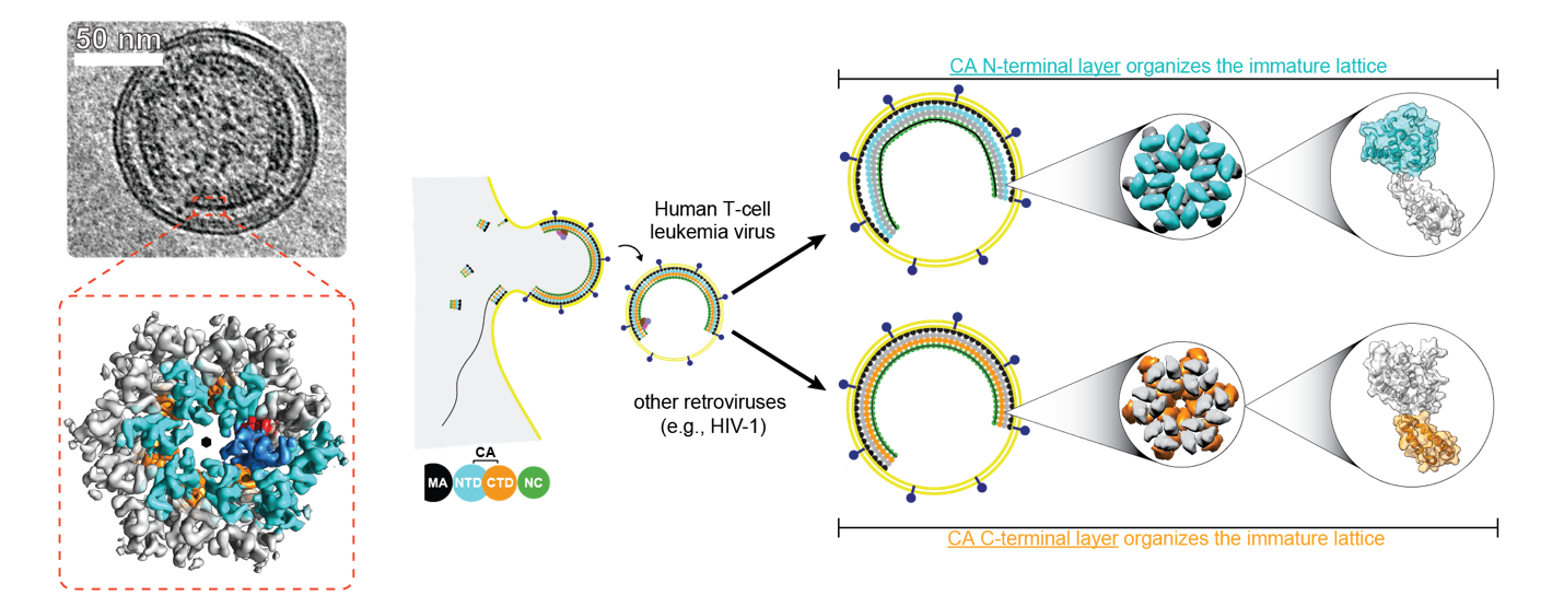
In Obr et al., (2024), we determined the structural organization of the immature HTLV-1 CA lattice. Retroviral CA proteins share a conserved tertiary fold, and prevailing models assume that lattice stabilization is mediated primarily through interactions of the C-terminal domain. Strikingly, our study revealed an unconventional assembly mechanism in HTLV-1, in which the N-terminal domain of CA mediates lattice stabilizing contacts. This finding provides a structural basis for the pronounced morphological plasticity observed in HTLV-1 particles, challenges the canonical paradigm of retroviral assembly, and demonstrates that distinct lattice architectures can arise from alternative usage of conserved structural elements. More broadly, this work highlights viral architecture as a key evolutionary variable and identifies new principles by which capsid stability and maturation can be regulated, beyond established HIV-1-centric models. The results were published in Obr M et al., Nature Structural and Molecular Biology (2024).

