
High-Resolution Optical Imaging for Biology
Biology

Johann Danzl
Professor
1 Career
- Since 2025 Professor, Institute of Science and Technology Austria (ISTA)
- 2017 – 2025 Assistant Professor, Institute of Science and Technology Austria (ISTA)
- 2017 Habilitation, biophysics (Priv. Doz.), Medical University Innsbruck, Innsbruck, Austria
- 2012 – 2016 Postdoc, Max Planck Institute for Biophys. Chem., Göttingen, Germany. Nanoscale fluorescence imaging, with Prof. Stefan W. Hell (Nobel Prize – Chemistry 2014)
- 2010 – 2011 Postdoc, Institute for Experimental Physics, Univ. of Innsbruck, Austria. Ultracold quantum gases, with Prof. Hanns-Christoph Nägerl
- 2010 PhD (Dr. rer. nat.), experimental quantum physics, Institute for Experimental Physics, Univ. of Innsbruck, Innsbruck, Austria. Advisor: Prof. H. C. Nägerl
- 2007 Diploma, experimental physics (Mag. rer. nat.); physics studies 1999 – 2007, Univ. of Innsbruck, Innsbruck, Austria
- 2005 Medical doctorate (Dr. med. univ., MD); medical studies 1997 – 2005, Medical Univ. Innsbruck, Innsbruck, Austria
1.1 Selected Distinctions
- 2026 The LICONN technology developed by the Danzl group was selected as one of seven “Technologies to watch in 2026” by Nature: https://www.nature.com/articles/d41586-026-00188-6
- 2025 “Golden Chalk Award” for best teaching at ISTA, awarded by AURAS, ISTA’s PhD student association
- 2024 FENS (Federation of European Neuroscience Societies) Forum 2024, Host Society Organizing Committee member, Vienna, Austria
- 2018 Otto-Kraupp Prize for best medical Habilitation in Austria, awarded by the Society of Physicians in Vienna (Gesellschaft der Ärzte in Wien), Austria
- 2010 Liechtenstein Prize 2009 awarded by the Principality of Liechtenstein, Vaduz, for the production of a quantum gas of deeply bound molecules
1.2 Selected Grants
- 2026 – 2030 ERC Advanced Grant, European Research Council, € 2.8 Mio.
Remark: This is the first ERC Advanced Grant at ISTA awarded to a professor who was hired as assistant professor and received tenure at ISTA - 2026 – 2027 Chan Zuckerberg Initiative Targeted Grant, $ 1.5 Mio.
- 2026 Unrestricted Gift by Google to support research in the Danzl group, $ 0.5 Mio.
1.3 Tech Transfer (Patents, Spin-Offs)
- 2024 XISTA (ISTA’s technology transfer subsidiary) filed a European Patent Application covering the invention of the expansion technology for LICONN (see research highlight for details; inventors: J. G. Danzl and ISTA PhD student M. Tavakoli). Patent application number EP24157341.9.
1.4 Selected Publications
- Light-microscopy based dense connectomic reconstruction of mammalian brain tissue;
M.R. Tavakoli, J. Lyudchik, M. Januszewski, V. Vistunou, N. Agudelo Dueñas, J. Vorlaufer, C. Sommer, C. Kreuzinger, B. Oliveira, A. Cenameri, G. Novarino, V. Jain, J.G. Danzl.
Nature (2025), DOI: 10.1038/s41586-025-08985-1. - Image-based 3D active sample stabilization on the nanometer scale for optical microscopy;
J. Vorlaufer, N. Semenov, C. Kreuzinger, M. G. Javoor, B. Zens, N. Agudelo Dueñas, M. R. Tavakoli, M. Šuplata, W. Jahr, J. Lyudchik, A. Wartak, F. Schur, J. G. Danzl.
Biophysical Reports (2025) DOI: https://doi.org/10.1016/j.bpr.2025.100211 - Super-resolution expansion microscopy in plant roots;
M. Gallei+, S. Truckenbrodt+, C. Kreuzinger, S. Inumella, V. Vistunou, C. Sommer, M.R. Tavakoli, N. Agudelo-Duenas, J. Vorlaufer, W. Jahr, M. Randuch, A. Johnson, E. Benkova, J. Friml, J.G. Danzl.
The Plant Cell (2025) https://doi.org/10.1093/plcell/koaf006 +Equal contrib. - Imaging brain tissue architecture across millimeter to nanometer scales;
J. Michalska, J. Lyudchik, P. Velicky, H. Stefanickova, J. F. Watson, A. Cenameri, C. Sommer, N. Amberg, A. Venturino, K. Roessler, T. Czech, R. Höftberger, S. Siegert, G. Novarino, P. Jonas, J.G. Danzl.
Nature Biotechnology (2023). https://www.nature.com/articles/s41587-023-01911-8. - Dense 4D nanoscale reconstruction of living brain tissue; P. Velicky, E. Miguel, J. M. Michalska, J. Lyudchik, D.Wei, Z. Lin, J. F. Watson, J. Troidl, J. Beyer, Y. Ben-Simon, C. Sommer, W. Jahr, A. Cenameri, J. Broichhagen, S. G. N. Grant, P. Jonas, G. Novarino, H. Pfister, B. Bickel and J. G. Danzl. Nature Methods (2023). https://www.nature.com/articles/s41592-023-01936-6.
- LIONESS, the technology presented in this paper, is one of the highlights in Nature Methods’ “Year in review 2023”: https://doi.org/10.1038/s41592-023-02158-6
2 Group Members
- Valentin Fonvieille, PhD Student since 2026
- Andrei Kalinichenko, PhD Student since 2026
- Liana Mukhametshina, PhD Student since 2024
- Vitali Vistunou, PhD Student since 2023
- Eder Miguel Villalba, Research Software Engineer since 2024
- Caroline Kreuzinger, Lab Manager/Technician since 2017
- Ivan Krylov, Research Technician since 2025
3 Research
Charting and mechanistically understanding biological systems depend on the technologies available to probe them. This is particularly evident in brain, where neurons form an incredibly dense, complex, and fine-grained arrangement that is challenging to analyze. A key aspiration of the group has been to make novel qualities of information accessible for biological tissues, integrating structural, molecular and functional characteristics. The optical imaging technologies we develop enable us and others to address biological questions in novel ways to better understand the brain in health and disease.
We take a fundamentally interdisciplinary approach, with physicists, computer scientists, biologists, and neuroscientists closely working together, also reflecting the PI’s dual background as MD and physicist. We have capitalized on the synergies across fields to address problems that cannot be tackled with a more narrow disciplinary approach.
To fully map the brain’s circuitry, the tissue’s architecture must be analyzed at a resolution that reveals the synaptic connections, i.e. its connectome, bridging from the tissue level to the nanometer scale. We have recently developed the first light microscopy technology for synapse-level connectomics (see “Research in Focus” section).
I see enormous continued potential for scientific innovation with integration across disciplines. Our technologies will be useful to map further organs, including in diseases such as cancer, and may eventually have translational impact for diagnostic and treatment decisions.
3.1 Current Scientific Projects/Directions
- Light-microscopy based connectomics
- Multi-modal mapping of brain tissue (structure, connectivity, molecular characteristics, function)
- 3D spatial transcriptomics in structural tissue context
- Analysis of human brain tissue across age, health and disease
- Integrated structural/molecular mapping of other organs
3.2 Research in Focus
To understand the brain’s functional architecture and its changes in disease, it’s necessary to chart the tissue, ultimately down to the level of the connectome, i.e. the arrangement of neurons and all their synaptic connections. This requires nanometer scale spatial resolution and has only been possible with electron microscopy (EM). EM reconstruction gives the structural detail required but is complex and fails to visualize the molecular characteristics of the cells. However, it’s the molecules, in particular proteins (and the associated transcripts) that define the functional repertoire of the cells. Light microscopy excels at visualizing specific molecules, but its resolution falls way short of the requirements for connectomics analysis, such that the interplay of structural characteristics and molecular mechanisms remain to a large degree elusive.
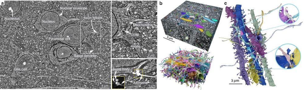
The group has recently developed “light microscopy based connectomics” (LICONN), the first light microscopy technology for dense reconstruction of brain tissue at synaptic resolution. LICONN serves as a platform to integrate different types of information (structure, connectivity, molecular characteristics, activity) in a straightforward manner. Rather than increasing microscope resolution, it uses a specifically engineered hydrogel embedding and expansion technology. This increases distances between cellular features 16-fold, yielding effective spatial resolution <20 nm even when imaging with standard off-the-shelf, high speed optical (spinning disc confocal) microscopes. LICONN enables automated segmentation using deep-learning methods, for which we have collaborated with the connectomics team at Google research.
LICONN was published in Nature in May 2025 (https://doi.org/10.1038/s41586-025-08985-1) and has been selected in January 2026 by Nature as one of seven “Technologies to watch in 2026” (https://www.nature.com/articles/d41586-026-00188-6). It has been adopted by leading institutions across the world and is currently transforming the field of connectomics. We expect that it will similarly be useful to better understand tissue function in health and disease in further organs, and potentially contribute to prediction of disease course or treatment response.

